Figure 10.
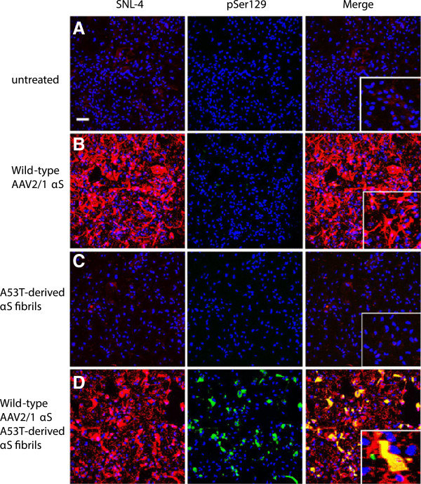
αS seeded inclusion formation in astrocyte cultures. Double immunofluorescence with SNL-4 (red) and pSer129 (green): (A) In untreated astrocyte cultures, there is very limited endogenous αS expression (SNL-4, red) and no hyperphosphorylated αS (pSer129, green). (B) rAAV2/1-mediated overexpression of human wild-type αS increases both the cell body and processes staining intensity for αS in the absence of hyperphosphorylated αS. (C) Addition of human A53T-derived αS fibrils alone showed similar endogenous expression levels of αS compared to the blank control along with the absence of hyperphosphorylated αS. (D) Both rAAV2/1-mediated overexpression of human wild-type αS and the addition of human A53T derived αS fibrils resulted in pSer129 immunoreactive inclusions in both the cell body and processes that overlap with SNL-4. Cultures were counterstained with DAPI (blue). Bar scale = 100 μm; insets = 25 μm.

