Figure 8.
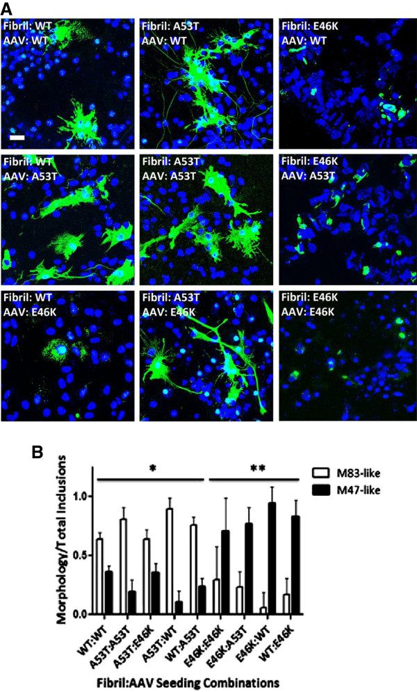
αS mutant specific and dominant morphological induction of αS aggregates. (A) Immunofluorescence analysis of morphological differences in αS aggregates is predominantly determined by the type of extracellular αS fibril challenge. Cultures were transduced with rAAV2/1 expressing wild-type, A53T, or E46K αS as indicated in each respective panel and described in Materials and Methods. Cultures were then treated with preformed fibrils comprised of wild-type, A53T, or E46K αS, as indicated in each respective panel. At one day post-seeding (and four days post-seeding for wild-type-wild-type), cultures were fixed and stained with pSer129 antibody (green) to assess for differences in morphology. Cells were counterstained with DAPI (blue). Bar scale = 25 μm. (B) Quantification of morphological differences for αS aggregates reveals statistical differences in inclusion morphology among seeding combinations. One-way ANOVA with Bonferroni correction for comparison of all groups to each other for flame-like M83 pathology reveals significant differences in inclusion morphology between all A53T fibrillar αS-seeded and E46K fibrillar αS-seeded (**) cultures, but not with WT and A53T combinations (*). Wild-type-wild-type combination (*) was more likely to result in M83-like inclusion morphology; however, when wild-type fibrils were used to seed either intracellularly expressed mutant proteins, the mutant protein predominantly determined inclusion morphology further indicating the dominant effect of the mutant proteins. p < 0.01; n = 6 fields/seeding combination. Values are means ± SD.

