Figure 4.
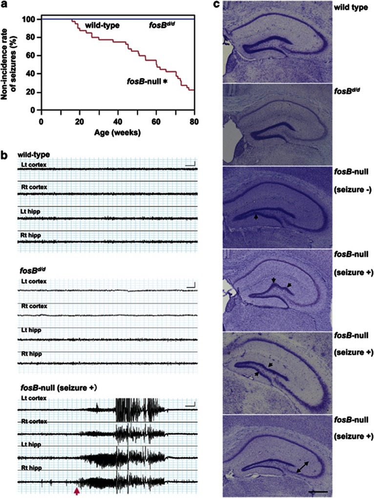
Aged fosB-null mice exhibit spontaneous seizures with epileptiform electroencephalograph discharges that originate in the hippocampus, and an abnormal DG structure. (a) The non-incidence rate of spontaneous seizures. Wild-type and fosBd/d mice, N=10; fosB-null mice, N=40. *p<0.0001, Kaplan–Meier method and log-rank test (χ2=40.94, df=2). (b) Concurrent hippocampal and cortical EEGs in 50- to 70-week wild-type (upper panel), fosBd/d (middle panel), and fosB-null mice (lower panel). Lt cortex, left cortex; Rt cortex, right cortex; Lt hipp, left hippocampus; Rt hipp, right hippocampus. N=5 in each group. Scale bars: X axis, 4 s; Y axis, 0.5 mV. (c) Nissl stained coronal hippocampal sections from wild-type, fosBd/d, and fosB-null mice. Characteristic abnormal arrangements (arrows) and thinning (bidirectional arrow) of the GCL are observed in fosB-null mice regardless of seizures. Seizure−, without any seizures; Seizure+, with seizures. Scale bar: 500 μm.

