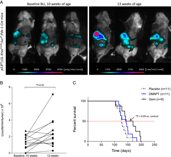
Figure 2.
Luciferase-expressing p53f/f; LSL-KrasG12D;lucl/l; Pdx-1-Cre mice. A) At 10 and 13 weeks of age, p53f/f; LSL-KrasG12D;lucl/l; Pdx-1-Cre mice were injected with D-luciferin (60 mg/kg; i.p.) and imaged to detect bioluminescence as shown for three representative mice. At each time point, a sequence of 15 images (2 minutes exposure time/image) was acquired. The image with the peak bioluminescence was used to assess relative change in photon fluence rate, e.g., counts/min in each voxel, using a lower threshold of 150 counts/min/sec (> 10 times background noise). The optical geometry was identical for all imaging sessions. B) Total photon flux (counts/min/tumor x 106) at 10 and 13 weeks of age (n = 14 mice). * P < 0.05 by two-tailed paired t-test. C) Median survival for each treatment group is shown in the Kaplan-Meier survival curve (placebo = 126 days; DMAPT = 143 days; Gem = 155 days). * P < 0.05 for gemcitabine vs. placebo by log-rank test.

