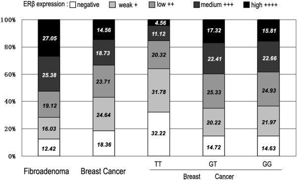
Figure 1.

Results of ERβ staining in five consecutive trials. Human breast cancer sections immunostained for ERβ were subdivided using a scoring system for ERβ expression into negative (no staining), weak (<20% staining), low (21–40% staining), medium (41–60% staining), and high (>61% staining) expression. The percentage of sections in each expression level is indicated. The first two columns showed the results for all the fibroadenoma (column 1) and breast cancer (column 2) sections. The last three columns showed the results for all breast cancer sections in each of the genotypes of rs1271572.
