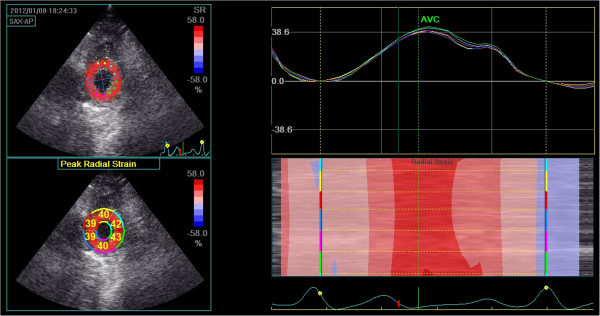
Figure 2.

Radial strain at the level of the apex. Top left: Peak systolic apical radial strain displayed using a colour map. Bottom left: Peak systolic apical radial strain displayed as a percentage for each individual segment. Top right: Left ventricular radial strain versus time curves corresponding to the 6 myocardial segments, Y-axis unit is %. Right bottom: Two-dimensional and M-curve colour-coded views show positive strain during systole.
