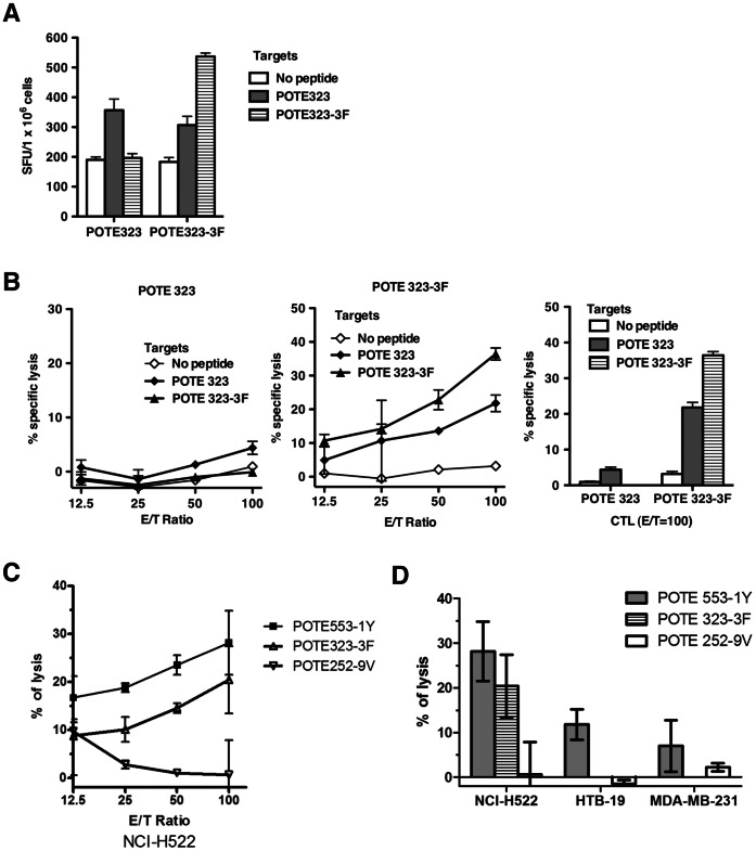
Figure 4. Immunogenicity of the wild-type and enhanced POTE323 epitopes and anti-tumor cytotoxicity induced by enhanced epitopes.
AAD mice were immunized s.c. with 50 nmol of peptide in 100 µl of emulsion as described in the Methods section. (A) Direct ex vivo IFN-γ ELISPOT assay using target cells pulsed with 1000 nM peptide. Figures show numbers of spots per million cells. (B) CTL cross-reactivity on POTE 323 and POTE 323-3F peptides. (C) Anti-tumor cytotoxicity against a POTE-expressing human lung cancer cell line NCI-H522 induced by POTE 553-1Y, 323-3F and 252-9V. HHD-2 mice were immunized with 50 nmol of the peptide indicated in 100 µl of emulsion and restimulated for 7 days with 1000 nM peptide before being tested for ability to lyse the tumor cells. (D) Summary of the anti-tumor cytotoxicity in POTE-expressing and non-POTE-expressing human tumor cells. NCI-H522 is a human lung cancer line expressing both POTE and HLA-A2, whereas HTB-19 is a human mammary carcinoma that expresses POTE but not HLA-A2, and MDA-MB-231 is a human mammary carcinoma cell line expressing HLA-A2 but not POTE. Killing of only the first of these shows the specificity for POTE in combination with HLA-A2. (Negative values are due to experimental 51Cr release slightly below spontaneous release with no effector cells, within experimental error, and so should be viewed as equivalent to zero).

