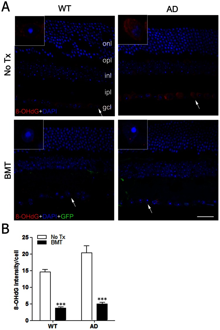
Figure 8. BMT results in reduced RGCL oxidative stress in aged wt and APPswe-PS1ΔE9 mice.
A: Immunofluorescence stains for 8-OHdG (red), an indicator of oxidative stress, are shown in representative retinal cross-sections from age-matched wt (left column) or APPswe-PS1ΔE9 (right column) mice that received no BMT transplant (top row) or BMT (bottom row). 8-OHdG immunofluorescence is primarily detected in RGCL neurons in non-transplanted wt and APPswe-PS1ΔE9 mice in a diffuse, perikaryal pattern. However, only focal, punctate immunostaining was observed in retinas from wt and APPswe-PS1ΔE9 mice that received BMT. Arrows indicate regions highlighted in insets. Scale bar = 20 μm. B: Quantification of 8-OHdG immunofluorescence relative intensity in RGCL neurons confirmed a significant reduction in 8-OHdG immunostaining in wt and APPswe-PS1ΔE9 mice that received BMT compared with non-transplanted controls, respectively. ***P<0.001, n = 6–10, two-way ANOVA followed by Bonferroni post test.

