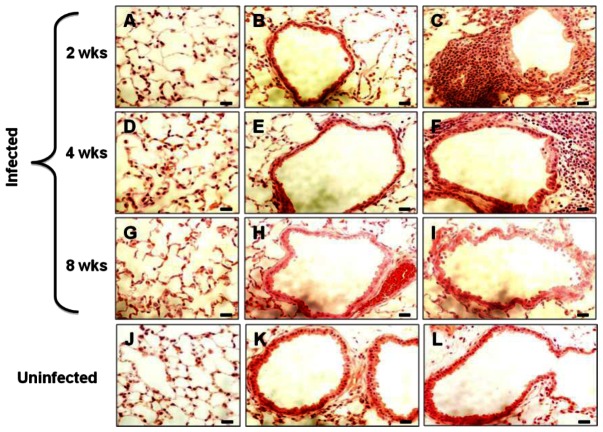
Figure 5. H&E stained lung sections of mice challenged with B. anthracis spores.
Lungs from infected and control mice were collected at 2, 4, and 8 weeks post-inoculation, fixed and subjected to H&E staining. Representative images displaying the alveoli and the airway from mice infected for 2 (A–C), 4 (D–F), and 8 weeks (G–I), and uninfected control mice (J–L) are shown. Scale bars represent 20 µm.

