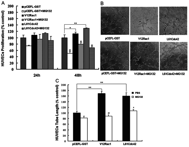
Figure 1. Effect of MG132 on HUVEC proliferation and tube formation induced by active Rac1/Cdc42.
The MTT and tube formation assays were performed as described in Material and Methods. (A) HUVECs were grown to confluence and were then cultured in preconditioned media (derived from GST–MCF-7, V12Rac1–MCF-7 and L61Cdc42–MCF-7 cells pretreated or not with MG132 for 12 h) for 24 h or 48 h; the GST–MCF-7 group was used as a control. (B) HUVECs were plated on Matrigel and incubated with the different preconditioned media for 6 h. Photographs were taken in five random power fields (200 ×). (C) Tube lengths were measured with Image-Pro Plus software. Histograms represent quantification of the HUVEC. The GST–MCF-7 group was used as a control. All data are expressed as mean ± standard deviation (SD) for three independent experiments. Statistical significance was assessed using one-way ANOVA and Student's t-test. *P<0.05, **P<0.01, for the GST–MCF-7 group vs the V12Rac1– or L61Cdc42– MCF-7 group. #P<0.05, ##P<0.01, for the MG132-added group vs the no-MG132 group.

