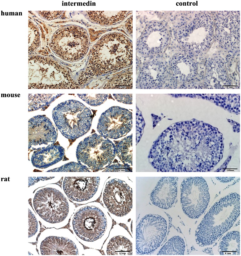
Figure 2. Immunolocalization of IMD in human, mouse and rat testis sections.
Intermedin positive immunostaining was located to both in the interstitial Leydig cells and to a lesser extent in the seminiferous tubules, including the tails of the spermatids. Representative sections of n = 4 stain with similar results; negative control omitting primary antibody for each section was presented in parallel at right panel; scale bars, 0.1 mm and 0.05 mm as indicated in each picture.

