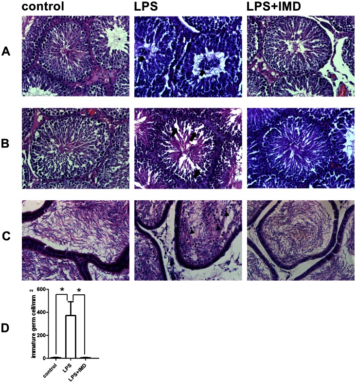
Figure 4. Histological examination of the rat testes and epididymis after LPS treatment or LPS and IMD cotreatment compared with saline control.
(A) LPS-treated testes 6 h after injection, showing accumulation of immature germ cells (indicated by arrows) in the seminiferous tubule lumen, which was not observed in the IMD-cotreated testis. (B) LPS-treated testes 72 h after injection, showing increased inter cellular gaps (indicated by arrows) due to disruption of cell-cell contract and/or loss of cells in the seminiferous epithelium, which was not observed in the IMD-cotreated testis. (C) LPS-treated epididymis 72 h after injection, showing large numbers of round immature germ cells (indicated by arrowheads) in the epididymal lumen, which was not observed in the IMD-cotreated epididymis. (D) Quantitive presentation of the detached round immature germ cells in the epididymal lumen by counting five random fields under 20X magnification. Data were expressed as mean ± SEM; * P<0.05 by Student t-test for (D); For (A), (B), and (C), representative sections of n = 5–7 rats with similar results.

