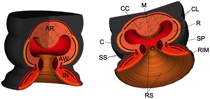
Figure 1. Schematic of the octopus suckers.

AR, acetabular roof; AW, acetabular wall; C, circular muscle (yellow sections); CC, cross connective tissue fibers (green crosses); CL, connective tissue layer; IN, infundibulum; M, meridional muscle (black lines); O, orifice; R, radial muscle (gray dotted line); RIM, rim around the infundibulum; RS, rough surface located on the surface of the infundibulum, orifice and acetabular protuberance; SP, primary sphincter muscle; SS, secondary sphincter muscle.
