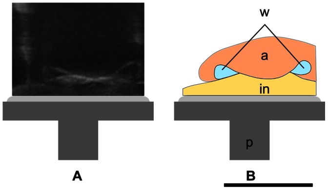
Figure 6. Octopus vulgaris sucker during adhesion process.

A) Ultrasonography of middle transverse section of sucker attached to the ultrasonographic probe. B) Schematic of A: a, acetabulum; in, infundibulum; w, water; p, ultrasonographic probe. The acetabular protuberance is in contact with the upper part of the side walls of the orifice. The scale bar equals 1 cm.
