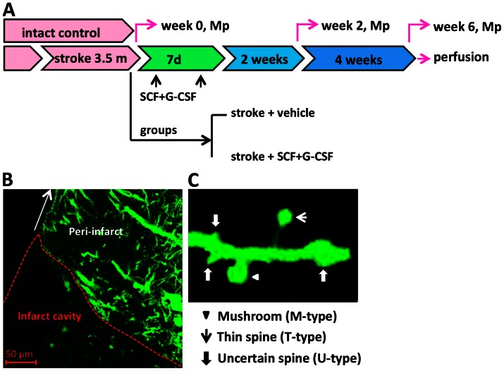
Figure 1. Experimental design and live brain imaging.
(A) Schematic diagram of experimental design of this study. At 3.5 months after induction of focal brain ischemia (stroke), mice were randomized to receive subcutaneous injection of vehicle or SCF+G-CSF for 7 days. Intact control mice received subcutaneous injection of vehicle for 7 days. The peri-infarct cortex was scanned through a thinned-skull window with a multiphoton microscope (Mp) before treatment (week 0), 2 and 6 weeks after treatment. At the end of the imaging, mice were transcardially perfused, and brains were removed and processed for immunohistochemistry. (B) A representative brain section from a thy-1-YFPH mouse with chronic stroke displays the peri-infarct cortex where the multiphoton imaging is performed. (C) A representative apical dendrite of the layer V pyramidal neuron expressing YFP illustrates different spine shapes including a mushroom type spine, a thin type spine and uncertain type spines. A mushroom type spine (M-type) with a large spine head and a thick neck. A thin type spine (T-type) with a small head and a narrow neck. Uncertain type spines (U-type) with undetectable heads and an equal diameter in the spine tip and stem or varicosity protuberance from the dendritic shaft.

