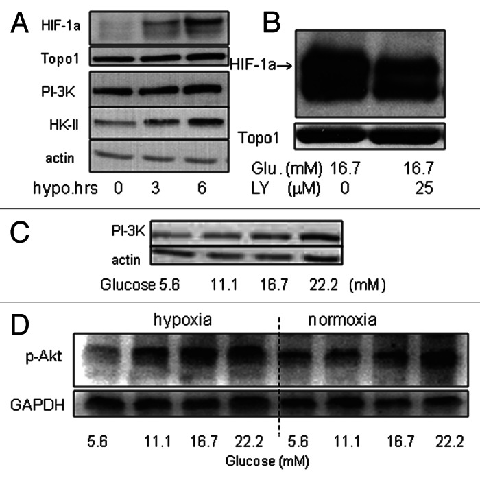
Figure 3.(A) HIF-1α, PI-3K and HK-II were determined by western blotting in wt-MiaPaCa2 cells after 0−6h hypoxic incubation. Topo1 and β-actin were used as loading controls. (B) Wt-MiaPaCa2 cells were incubated in hypoxia for six hours, using culture media that contained 16.7mM glucose in the absence or presence of LY294002 (LY, 25 μM). HIF-1α was determined by western blotting. (C) Wt-MiaPaCa2 cells underwent 6 h hypoxic incubation with different amounts of glucose. The p85 subunit of PI-3K was determined by western blotting. (D) Wt-MiaPaCa2 cells were incubated with different amounts of glucose in hypoxia or normoxia. Phospho-Akt was determined by western blotting, using GAPDH as a loading control.
