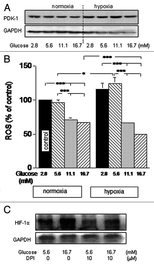
Figure 6. Wt-MiaPaCa2 cells were incubated with different amounts of glucose in normoxia or hypoxia for six hours. (A) PDK-1 was determined by western blotting, using GAPDH as a loading control. (B) ROS were determined by flow cytometry. n = 9. * p < 0.05 and *** p < 0.001. (C) Hypoxic wt-MiaPaCa2 cells were incubated in the absence or presence of diphenyleneiodonium (DPI, 10 μM). HIF-1α was determined by western blotting.
