
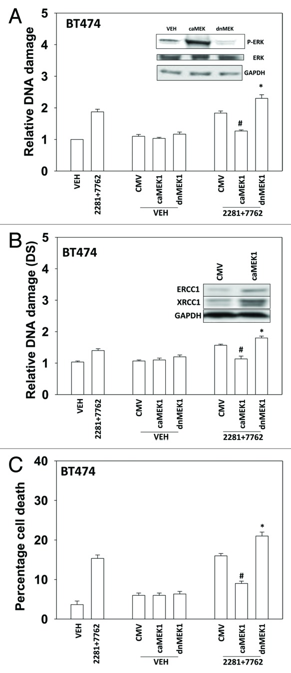
Figure 6. MER-ERK signaling regulates the DNA damage response following PARP1 and CHK1 inhibitor treatment. (A) BT474 cells were infected with empty vector adenovirus (CMV) or viruses to express dominant negative MEK1 (dnMEK1) or activated MEK1 (caMEK1). Twenty four h after infection cells were treated with AZD2281 (1 μM) and AZD7762 (25 nM) in combination for 24h. Cells were isolated and subjected to alkaline comet assay. The length of the tail being scored 1–5 (n = 3 ± SEM). * p < 0.05 value greater than corresponding vehicle control; # p < 0.05 value less than corresponding vehicle control. Inset blot: the levels of ERK1/2 phosphorylation in cells expressing caMEK1 and dnMEK1. (B) BT474 cells were infected with empty vector adenovirus (CMV) or viruses to express dominant negative MEK1 (dnMEK1) or activated MEK1 (caMEK1). Twenty four h after infection cells were treated with AZD2281 (1 μM) and AZD7762 (25 nM) in combination for 24h. Cells were isolated and subjected to neutral comet assay. The length of the tail being scored 1–5 (n = 3 ± SEM). * p < 0.05 value greater than corresponding vehicle control; # p < 0.05 value less than corresponding vehicle control. Inset blot: the levels of ERCC1 and XRCC1 in cells expressing caMEK1. (C) BT474 cells were infected with empty vector adenovirus (CMV) or viruses to express dominant negative MEK1 (dnMEK1) or activated MEK1 (caMEK1). Twenty four h after infection cells were treated with AZD2281 (1 μM) and AZD7762 (25 nM) in combination. Cells were isolated 24h after exposure, and viability was determined using trypan blue exclusion. (n = 3 ± SEM). * p < 0.05 value greater than corresponding vehicle control; # p < 0.05 value less than corresponding vehicle control.