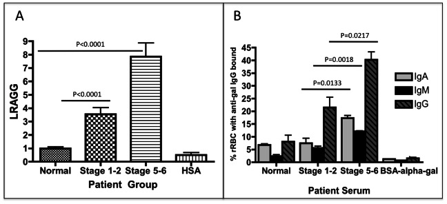
Figure 2. Increased lectin reactive anti-gal IgG from patients with increasing levels of liver fibrosis.

A) Compared to commercially purchased normal human sera, patients with limited liver fibrosis (stage 1–2) have a 3.5 (±1.4) fold increase in lectin reactive anti-gal IgG (LRAGG). More advanced (Stage 5–6) fibrosis patients have a 7.9 (±2.4) fold increase in LRAGG. The fold increase in LRAGG is statistically significant (P<0.001). When serum from more advanced fibrotic patients is used on plates coated with HSA alone and not HSA-alpha-gal, no signal is observed (HSA lane). B) The level of anti-gal IgA, IgM and IgG bound to target rabbit red blood cells as a function of liver fibrosis. There is a statistically significant increase in anti-gal IgA from commercially purchased normal human serum to serum from patients with advanced fibrosis (P = 0.0048); Anti-gal IgA also significantly increases from limited to advanced fibrosis (P = 0.0133). Anti-gal IgM significantly increases from control to limited fibrosis (P = 0.02) and from control to advanced fibrosis (P = 0.002); there is also a significant increase in anti-gal IgM from limited to advanced fibrosis (P = 0.0018). Anti-gal IgG is significantly elevated in advanced fibrosis compared to control (P = 0.0075) and also from limited to advanced fibrosis (P = 0.0217). 50 mM of BSA-alpha-gal can prevent binding of antibodies to RBCC. See text for more details. For panels A & B, samples size is Normal, n = 21; Stage 1–2, n = 22; and Cirrhosis, n = 39.
