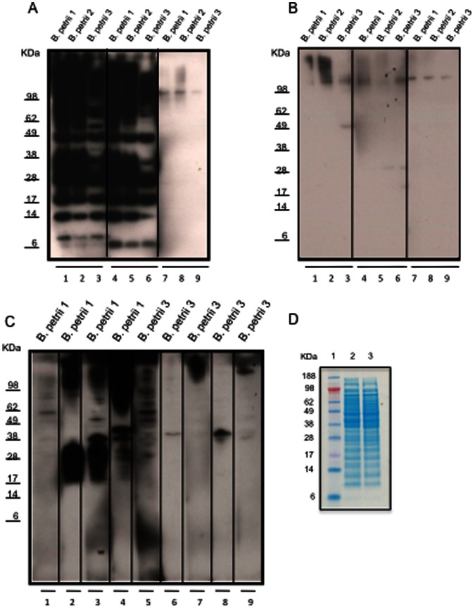
Figure 3. Immunoblots with sera from mice inoculated with B. petrii 1 extracts (A), B. petrii 3 extracts (B) or live B. petrii 1 or B. petrii 3 (C) and SDS–PAGE analysis of B. petrii 1 and B. petrii 3 extracts (D).
An aliquot containing 10 µg of bacterial proteins was electrophoresed on 12% SDS–PAGE, and then transferred to PVDF membrane. The membrane was incubated with sera from mice (1∶200 dilution) previously inoculated with extracts from B. petrii 1 (3A lanes 1–3 and 4–6 for two different mice) or B. petrii 3 (3B lanes 1–3 and 4–6 for two different mice) or with live B. petrii 1 (3C lanes 2, 3 and 4 for three different mice) or live B. petrii 3 (3C lanes 6, 7, 8 and 9 for four different mice). Uninoculated normal mouse sera controls are shown in 3A lanes 7–9, 3B lanes 7–9 and 3C lanes 1 and 5. The membrane was then incubated with horseradish peroxidase conjugated sheep anti-mouse IgG (1∶10000) and blots developed using the enhanced chemiluminescence kit. (D) B. petrii 1 and B. petrii 3 protein extracts were analyzed by SDS–PAGE (Invitrogen NuPAGE 12% Bis-Tris gel) and stained with Coomassie Blue R-250 (Invitrogen). Lane 1 Standard (SeeBlue Plus2 pre-stained Standard Invitrogen); lanes 2, B. petrii 1 extracts; and lane 3, B. petrii 3 extracts (10 µg proteins per lane).

