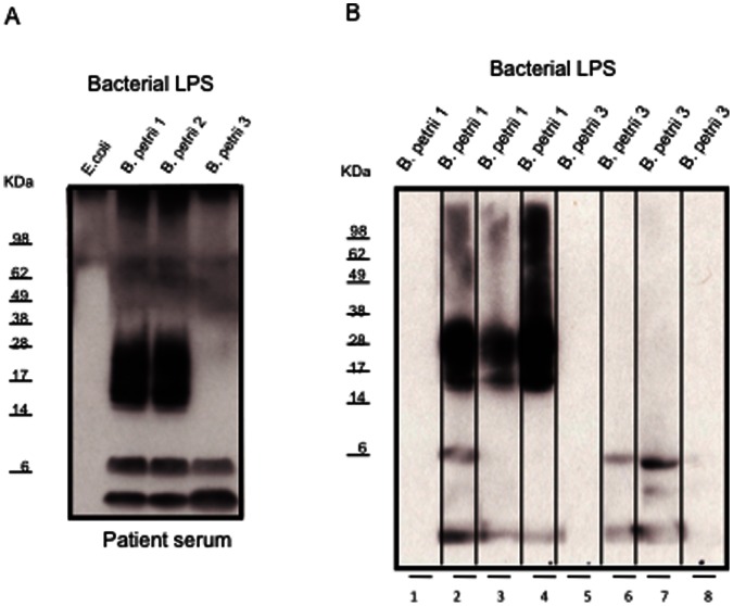
Figure 4. Immunoblots with patient (A) or mice (B) sera against B. petrii LPS.
B. petrii LPS was isolated from bacterial pellets by using a LPS extraction kit. B. petrii LPS (10 µg/lane) or commercially obtained E. coli LPS control (3 µg/lane) were electrophoresed on 12% SDS–PAGE, and then transferred to PVDF membrane. The membrane was incubated with the patient serum (1∶5000 dilution) or sera from mice (1∶200 dilution) previously inoculated with B. petrii. B. petrii 1 LPS (4B lanes 2, 3 and 4 for three mice) or B. petrii 3 LPS (4B lanes 6, 7 and 8 for three mice) or with uninoculated normal mouse sera controls (4B lanes 1 and 5). The membrane was then incubated with horseradish peroxidase conjugated sheep anti-human (A) or anti-mice (B) IgG (1∶10000) and blots developed using the enhanced chemiluminescence kit.

