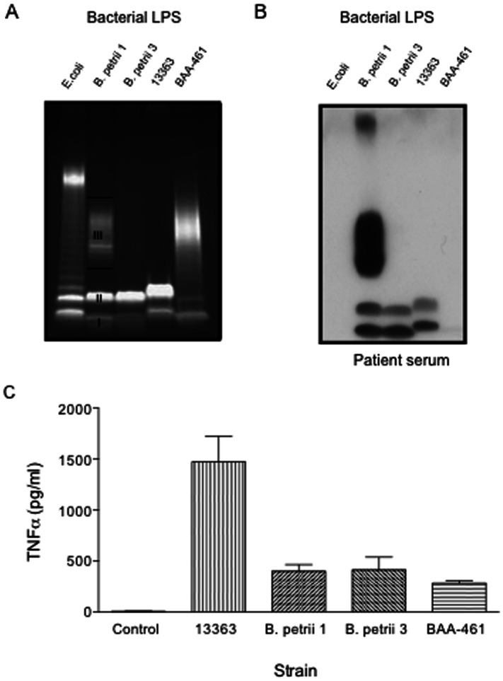
Figure 5. SDS PAGE (A), immunoblots with our patient’s serum (B) and TNF-α induction of B. petrii LPS.
(A) Five ml overnight cultures from each bacterial strain (B. petrii 1, B. petrii 3, NCTC 13363 and ATCC BAA-461) were spun 10 min at 8000g to harvest bacterial cells and LPS isolated by using a LPS extraction kit. Extracted LPS samples were separated on a 12% SDS-PAGE and stained using Pro-Q Emerald 300 Lipopolysaccharide gel stain kit. LPS concentrations were measured spectrophotometrically at 205 nm. Control E coli LPS was obtained commercially. I, II and III indicate LPS bands I, II and III respectively, described in the text (B) Immunoblots of our patient’s serum against LPS were performed as described in Fig. 4A. (C) Purified LPS (200 ng/ml) was added to human peripheral blood mononuclear cells (PBMCs) from normal volunteers, and supernatants were collected for cytokine measurements after 20 hours. p<0.001 for NCTC 13363 vs B. petrii 1, B. petrii 3 or BAA-461. Graph shows mean and SEM from three experiments.

