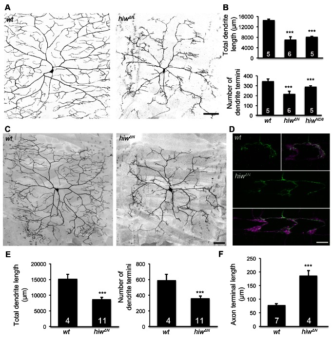
Figure 1. Hiw differentially regulates dendrite and axon growth in C4da neurons.
(A) Dendrites of the C4da neuron ddaC in hiwΔN homozygous mutant larvae are reduced, as compared to wild-type (wt). C4da neurons were labeled by the C4da marker ppk-CD4::tdTomato. Scale bar, 100 µm. (B) Bar charts showing the quantification of total dendrite length (top), number of dendrite termini (bottom) of ddaC in wt, hiwΔN, and hiwND8 larvae. Sample numbers are shown in the bars of the bar charts throughout this article. (C–D) hiw mutant MARCM clones exhibit impaired dendritic growth and overgrowth of axon terminals. (C) Representative dendrites of wt and hiwΔN mutant ddaC neurons. Scale bar, 50 µm. (D) Representative axon terminals of a single wt ddaC and a single hiwΔN mutant ddaC. The axon terminals of wild-type ddaC clones (green) extend within one segment length of the C4da neuropil (magenta) labeled by ppk-CD4::tdTomato. The axon terminals of hiwΔN mutant clones (green) expand over multiple segment lengths of the C4da neuropil (magenta). Scale bar, 10 µm. (E) Quantification of total dendrite length (left) and number of dendrite termini (right) of wt and hiwΔN MARCM clones. (F) Quantification of axon terminal length of wt and hiwΔN MARCM clones.

