
Figure 3.
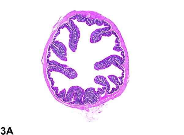
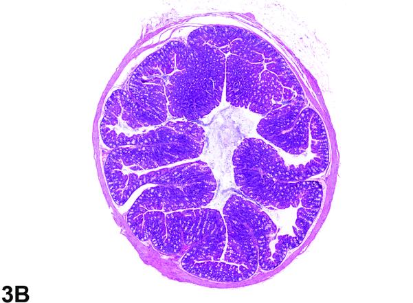
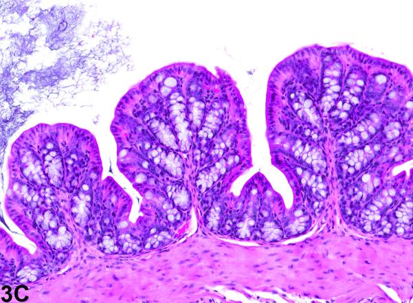
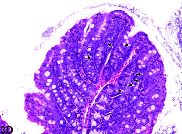
Colon from the 40-week study of senna. A, a cross section of the colon from a control male mouse. 3X, H&E. B, a cross section of the colon from a male F1 p53+/− mouse exposed to 10,000 ppm. There is moderate epithelial hyperplasia, with diffuse thickening of the epithelium. 3X, H&E. C, Higher magnification of colonic epithelium from a control male mouse. 20X, H&E. D, a section of colon from a male mouse exposed to 10,000 ppm senna. The epithelium is thickened as evidenced by increased distance from the luminal surface to the base of the crypts. Epithelial cells are increased in number and are basophilic and crowded, the cells lack the orderly arrangement seen in the control mouse, and there are numerous mitotic figures (arrows). 20X, H&E.