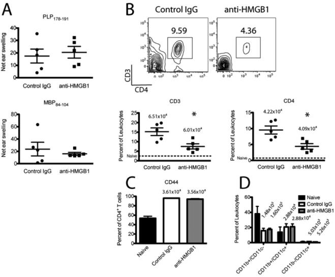
Figure 4. HMGB1 neutralization results in sustained inhibition of CNS T cell infiltration/expansion.
R-EAE was induced in SJL/J mice by immunization with 50 μg PLP139-151/CFA s.c., and 100 μg anti-HMGB1 neutralizing antibody or isotype control antibody was injected i.v. at disease remission (day 19 p.i.). PLP178-191- and MBP84-104-specific CD4+ T cell responses were assessed by DTH assays 75 days p.i. (A). Spinal cords were harvested for flow cytometric analysis at the third relapse (78 days p.i.), dissociated, and single cells were purified. T cells were stained for CD3, CD4, and CD44 expression (B,C), and APCs were stained for CD11b and CD11c expression (D). Data are representative of two experiments of ≥ 5 mice per group. Asterisks denote a significant difference in DTH responses or flow cytometric quantification between treatment groups by Student's t-tests (p < 0.05).

