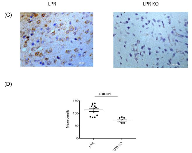
Figure 6. Fn14 deficiency decreases expression of RANTES, C3 and CXCL11 in the brain of MRL/lpr mice.
(A) The gene expression of RANTES, C3 and CXCL11 in the cortex of 19 week old, female and male MRL/lpr Fn14WT and Fn14KO mice was measured by real-time PCR, and normalized to GAPDH. For the PCR, fold changes were calculated in reference to the MRL/lpr Fn14KO mice, whose mean value was set at 1. (B) The gene expression of RANTES, C3 and CXCL11 in the whole brain of 40 week old, female and male MRL/lpr Fn14WT and Fn14KO mice was measured by real-time PCR, and normalized to GAPDH. For the PCR, fold changes were calculated in reference to the MRL/lpr Fn14KO mice, whose mean value was set at 1. (C) Representative images of the immunohistochemical staining for RANTES in the frontal cortex of female MRL/lpr Fn14WT and Fn14KO mice at 40 weeks of age. Sections were examined using a Zeiss AxioObserver at 40X magnification. (D) RANTES staining quantitation was performed by Image J software. Staining intensity in 10 randomly selected areas was quantitated in each section, and the average of the density for each section was then calculated. Female mice, full dots; Male mice, open dots.


