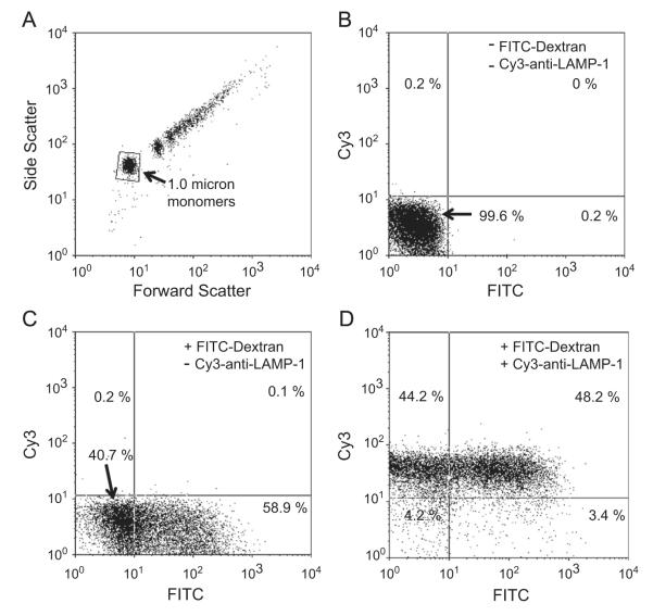
Fig. 1.
Permeability properties of magnetically isolated neutrophil phagosomes as assessed by flow cytometry. Phagosomes were isolated from human neutrophils 15 min after phagocytosis of opsonized 1-μm paramagnetic beads in the (C and D) presence or (B) absence of FITC-labeled dextran as described under Materials and methods. (D) The isolated phagosomes were stained with Cy3-conjugated rabbit anti-LAMP-1 as a marker for mature phagosomes and subjected to two-color analysis by flow cytometry. (A) The forward and side light-scatter properties of the preparation. A typical scattering pattern consisted of a monomeric fraction (arrow) and a series of higher order aggregates. Analysis of the fluorescence properties of the monomeric fraction, as gated, is shown in (B–D). (B) Phagosomes prepared in the absence of FITC–dextran and Cy3–anti-LAMP-1. Less than 0.2% of the population was positive for either marker. (C) When FITC–dextran was included in the incubation medium during particle phagocytosis, 58.9% of the phagosomes retained the FITC label after lysis and isolation. (D) When this preparation was labeled with Cy3–anti-LAMP-1 48.2% of the phagosome population was positive for LAMP-1 and FITC–dextran.

