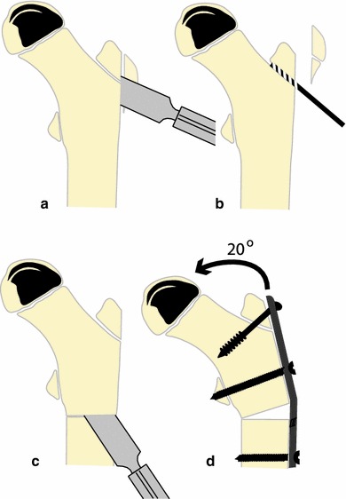
Fig. 1.

The technique of surgical containment used in this study. A sliver of the bony flare of the greater trochanter is removed (a), the trochanteric growth plate is drilled (b), a subtrochanteric osteotomy is performed (c) and the fragments are fixed with a pre-bent plate to ensure a varus angulation of 20° (d)
