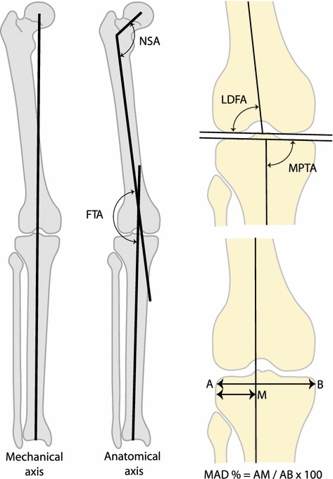
Fig. 2.

The different angles measured on full-length standing radiographs included the neck-shaft angle (NSA), the femur-tibial angle (FTA), the lateral distal femoral angle (LDFA) and the medial proximal tibial angle (MPTA). The mechanical axis deviation was expressed as a ratio of the distance of the axis from the lateral border of the tibia to the width of the tibial articular surface. A value less than 50 % indicates a valgus alignment of the knee while a value greater than 50 % indicates a varus alignment
