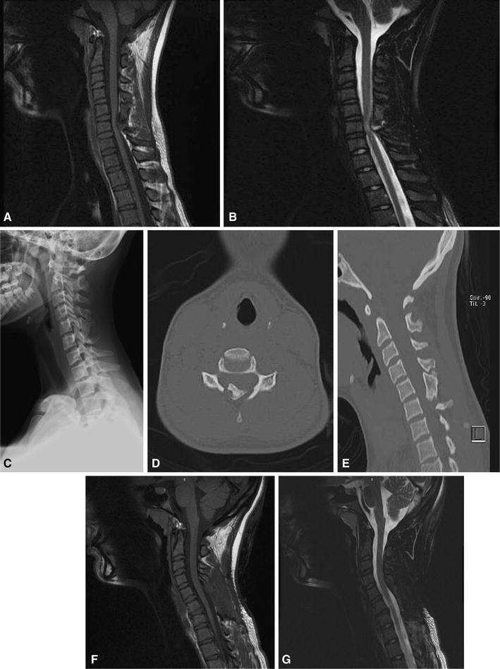
Fig. 1.

A 15-year-old female with multiple hereditary exostoses (MHE) presented with hip and posterior leg pain. Magnetic resonance imaging (MRI) screening of the entire spine was undertaken, given concern for radicular-type symptoms. a, b Sagittal T1 and T2 imaging of the cervical spine revealed a large posteriorly based osteochondroma compressing the spinal cord, with signal change on T2 imaging. c The lesion is not evident in plain radiographs. d, e Computed tomography (CT) underestimates the size of the lesion, given the large cartilage cap. Nevertheless, the appearance of the osteochondroma on computed tomography (CT) is concerning for spinal stenosis. This patient underwent surgical decompression, with no improvement in her hip and leg symptoms. f, g Sagittal T1 and T2 imaging of the cervical spine status post laminectomy. Note the resolution of the previous signal change on T2
