Figure 1.
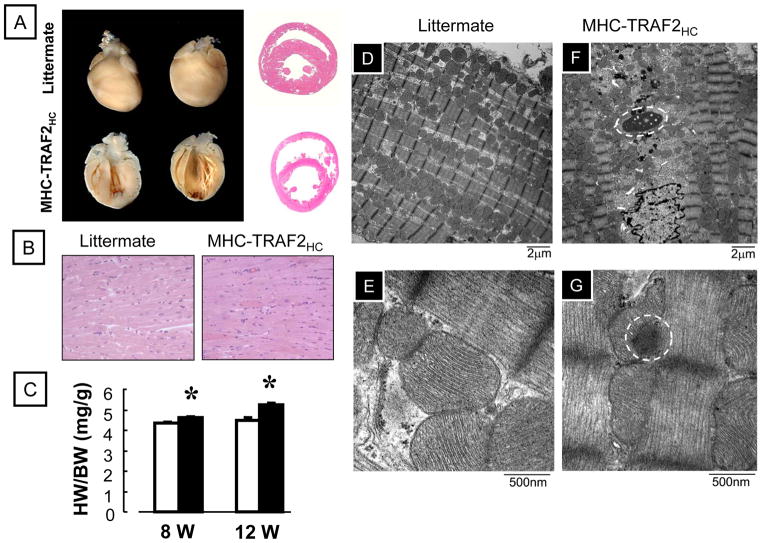
Characterization of MHC-TRAF2HC transgenic mice. (A) Photographs of whole hearts, coronal and sagittal sections of both LM and MHC-TRAF2HC mouse hearts (12 weeks). (B) Representative hematoxylin-eosin stained cross sections at the level of the papillary muscles (400x). (C) Heart-weight-to-body-weight ratio (mg/g) of LM and MHC-TRAF2HC at 8 and 12 weeks (n= 6–8 mice/group/time point) (*= p< 0.05 vs. LM at the indicated time point) (D–G) Representative transmission electron micrographs from 12 week MHC-TRAF2HC transgenic mice and LM at low (x7500, D,F) and high (x 60,000, E,G) magnification. Protein aggregates are enclosed by the circles.

