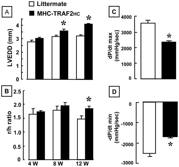
Figure 2.
LV structure and function. (A) 2-D echocardiographic assessment of left-ventricular end diastolic dimension (LVEDD) and (B) r/h (radius/wall thickness) ratio of 12 week LM and MHC-TRAF2HC at 4, 8 and 12 weeks (n= 6–8 mice/group/time point). (*= p< 0.05 vs. LM at the indicated time point) (C) LV +dP/dt and (D) LV −dP/dt assessed in 12 week in LM and MHC-TRAF2HC (n= 6 mice/group) (*= p< 0.05 vs. LM at 12 weeks).

