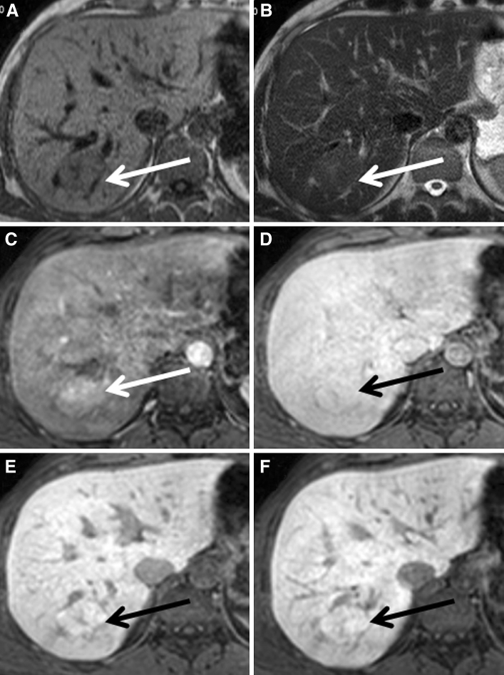
Fig. 2.
A patient presenting with a typical FNH on conventional FNH, which appeared homogeneously hyperintense to the surrounding parenchyma during hepatobiliary phases. A–F The FNH is visible on T1, T2, and during arterial phase, portal-venous phase, and 5 and 10 min hepatobiliary phase, respectively (white and black arrows).

