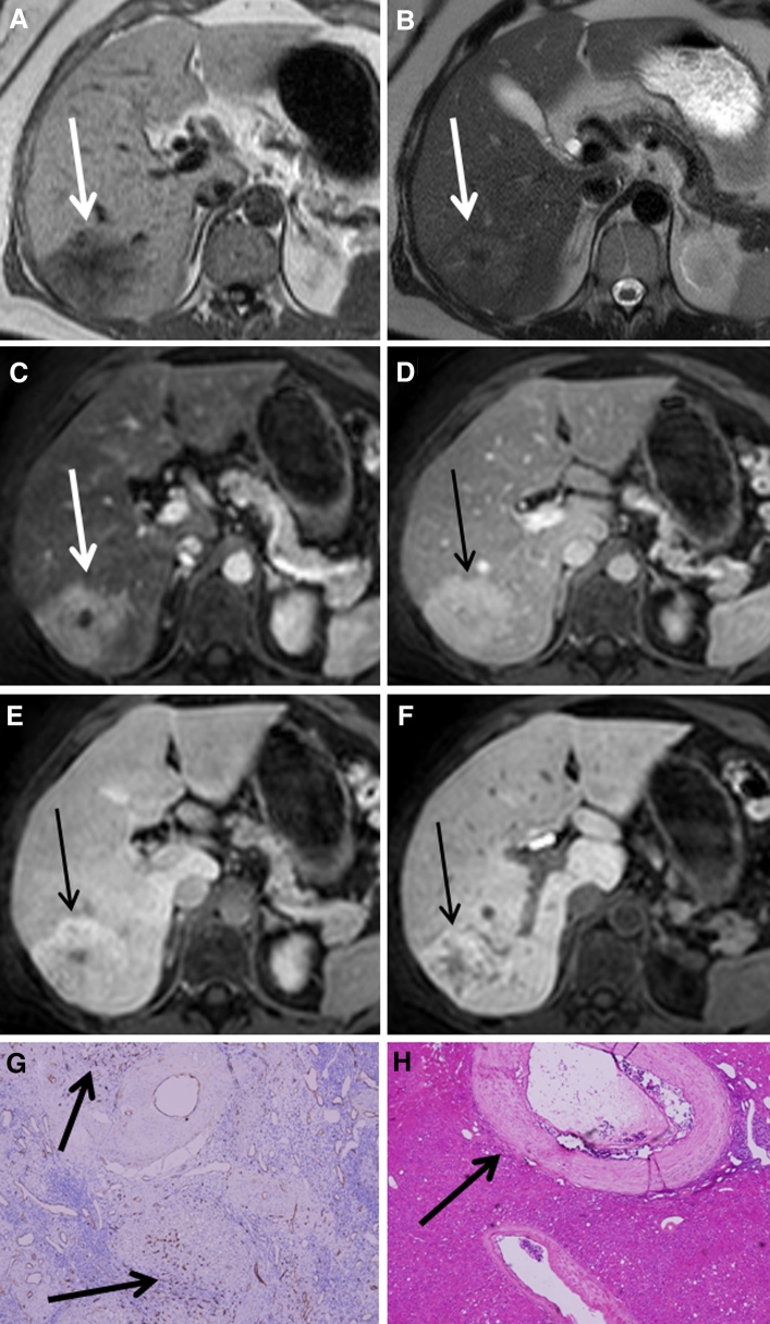
Fig. 3.
A patient with a histologically proven FNH presenting as an inhomogenously hyperintense lesion on hepatobiliary phases (Tumor C). A–F The FNH is visible on T1, T2, and during arterial phase, portal-venous phase, and 5 and 10 min hepatobiliary phase, respectively (white and black arrows). Initially, the lesion presents as a hyperintense lesion with a non-enhancing central scar during arterial phase, but over time the lesion characteristics change resulting in an inhomogeneous appearance during hepatobiliary phase. G CD34 immunohistochemistry shows diffuse positivity throughout the lesion (brown staining at arrows). H Hematoxylin and eosin staining showing an abnormal, enlarged vessel with thickened vessel wall (arrow). The inhomogeneous appearance on hepatobiliary phase is presumably related to areas with ischemic injury and ductular metaplasia due to vascular abnormalities, which are alternated by areas with ductular proliferation.

