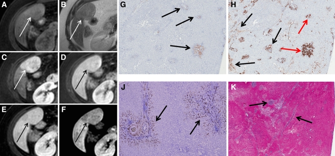
Fig. 6.
Another patient presenting with a hypo-intense type FNH in segment 5 (Tumor B). A–F The FNH is visible on T1, T2, and during arterial phase, portal-venous phase, and 5 and 10 min hepatobiliary phase, respectively (white and black arrows). The FNH is visible during all phases and initially presents as a hypervascular lesion on arterial phase without a central scar. During hepatobiliary phases, the lesion signal is less intense compared to the surrounding parenchyma, resulting in a slightly hypointense aspect. G CK 19 immunohistochemistry showing weakly CK 19 positive ductular proliferation in the lesion periphery (arrows), while the lesion centre is almost completely negative. H CK7 immunohistochemistry shows diffuse positivity in the lesion representing both ductular proliferation and ductular metaplasia (black arrows); the bile-ducts that are positive for CK19 (G) are positive for CK7 as well (red arrows). The ductular metaplasia is more pronounced than the ductular proliferation and is visible both in the lesion centre as in the lesion periphery. J CD34 immunohistochemistry shows expression around the fibrous tissue (arrows). K Azan staining demonstrates the presence of fibrous tissue throughout the lesion (black arrows pointing out blue areas), although a central scar cannot be identified.

