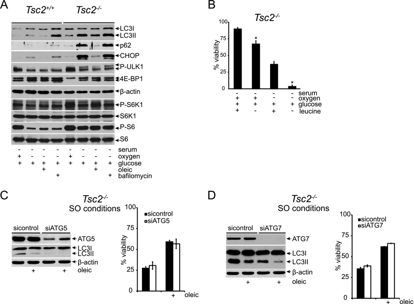
Figure 4.
Restoration of autophagic flux fails to rescue Tsc2−/−, p53−/− cell viability under SO conditions. (A) Autophagic signaling and flux were examined in Tsc2+/+, p53−/− and Tsc2−/−, p53−/− MEFs under S and SO conditions for 24 h by assaying the levels of LC3I, LC3II, p62, and CHOP with and without 100 nM bafilomycin. mTORC1 signaling was monitored by assessing the phosphorylation status of ULK1, 4E-BP1, S6K1, and S6. (B) The effect of leucine deprivation on viability under 48 h of S and SO conditions was examined (P < 0.001). (C,D) The contribution of autophagy to oleic acid rescue of Tsc2−/−, p53−/− MEF cell death under SO conditions was assayed. Pools of Tsc2−/−, p53−/− MEFs were depleted of ATG5 (C) or ATG7 (D) protein using siRNAs and cultured under SO conditions in the presence and absence of oleic acid. After 48 h, viability was assessed by flow cytometry. The degree of knockdown was determined by Western blot.

