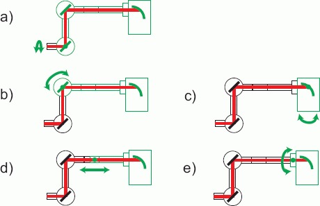
Figure 4.

(online color at: www.biophotonics-journal.org) Possible rotations and elongations of the gantry leading to many degrees of freedom for patient treatment (“gantry scanning”). Black parts do not move, while green parts can move. Subfigure (a) shows a normal gantry movement whereas (b) and (c) demonstrate a tilt at different parts of the gantry. In (d) an elongation is illustrated and (e) represents a rotation of the treatment head around the beam axis.
