
Figure 3.
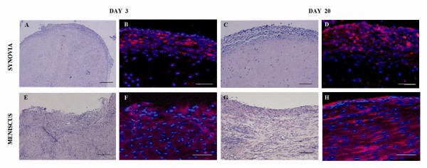
Biodistribution of CM-Dil labeled ASCs. Detection of labeled-ASCs in synovial lining layer (A-D) and medial meniscus (E-H) of representative specimens at 3 and 20 days from ASC administration under bright field (A, C, E, G) and epi-fluorescence (B, D, F, H). Blue staining = nuclei; Red staining = CM-Dil labeled-ASCs. Scale bars = 500 μm.