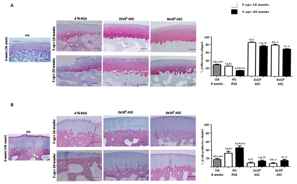
Figure 8.

Immunohistochemical analysis for collagen II and I in cartilage. Photomicrographs of representative specimens evaluated for type II (A) and I (B) collagens in medial femoral condyle of OA, 4% RSA and ASC-treated groups. Data are reported as mean ± SD. Scale bars = 100 μm. Statistical values of at least P < 0.01 were observed in all the comparisons.
