
Figure 7.
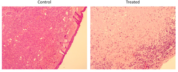
H&E slides of untreated and treated positive surgical margins. Positive-margin controls (left) show malignant cells with large nuclei and irregular chromatin. Treated positive margins H&E slides (right) represent tissue margins four weeks after treatment with GLV-1h153. Large areas of fibrosis and necrosis can be seen without any viable cancer cells.