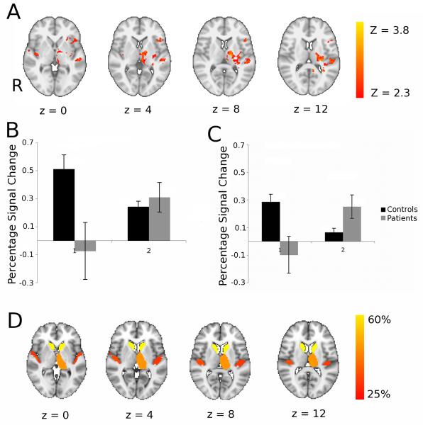
Figure 4. Patients showed increased brain activation with motor practice relative to controls and many of these brain regions correspond well with those that had impaired white matter connectivity at baseline.
A. Brain regions showing significant differences between patients and controls for task performance at baseline versus performance following three weeks of task practice. Axial slices at MNI levels indicated were thresholded at an initial cluster forming threshold of Z>2.3, and a corrected cluster extent threshold of p<0.05. The left hand side of the brain (ipsilesional in patients) is displayed on the right hand side of each brain slice. See Table 3 for details of local maxima. B,C: Region of interest analyses to characterise direction of activation change in clusters showing group by time interaction in the thalamus (B) and insula (C). D. Brain atlas regions with reduced white matter connectivity at baseline in patients relative to healthy controls. To identify these, probabilistic diffusion MRI tractography was performed and mean probability of connectivity to other brain regions were measured for each atlas region. The colour scheme indicates % reduction in connectivity probability in patients compared to controls and all regions showing 25% or more reduction are coloured.

