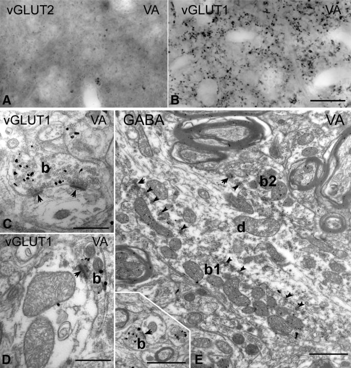
Figure 5.
Thalamic territories without large vGLUT1 or vGLUT2 terminals. A, B, High-power light microscopic images of vGLUT2-immunostained (A) and vGLUT1-immunostained (B) sections from the ventral anterior nucleus display the lack of large terminal labeling by both markers. C, D, At the electron microscopic level, vGLUT1-positive terminals show the features of RS-type terminals. Most of them form a single synapse or more rarely two synapses and contained a maximum of two mitochondria. E, The large terminals of the ventral anterior nucleus (b1 and b2) are negative for vGLUT1 but show GABA immunoreactivity after postembedding GABA immunogold staining. Inset shows a vGLUT1-immunoreactive terminal at the same magnification. Scale bars: A, B, 20 μm; C, D, 500 nm; E and inset, 1 μm.

