
Figure 8.
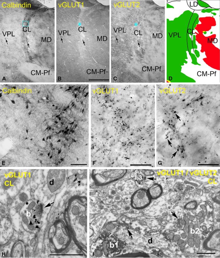
Excitatory inputs of the rostral intralaminar nuclei. A–D, Delineation of the intralaminar nuclei. Low-power light micrographs of adjacent sections immunostained for calbindin (A), vGLUT1 (B), and vGLUT2 (C). Note that the borders of the intralaminar nuclei are discernible only in the calbindin immunostaining. Arrows indicate corresponding capillaries. The color map (D) indicate the distribution of large terminals (green, large vGLUT2 terminals; red, large vGLUT1 terminals). Intralaminar nuclei are continuous laterally with VPL but contain no large vGLUT1 terminals, unlike the mediodorsal nucleus. E–G, High-power light microscopic images of the blue boxed regions in A–C, respectively. Note the lack of large vGLUT1-positive terminals and medium-sized vGLUT2 terminals (arrows in G) in the centrolateral nucleus. H, At the electron microscopic level, a vGLUT1-positive terminal (b) displays RS features. I, After double immunostaining, the size difference between RL-type vGLUT2 terminals (b1, b2, DAB precipitate) and RS-type vGLUT1-immunoreactive terminals (b3, silver-intensified gold) are apparent. Scale bars: A–C, 2 mm; E, 100 μm; F, G, 20 μm; H, I, 1 μm.