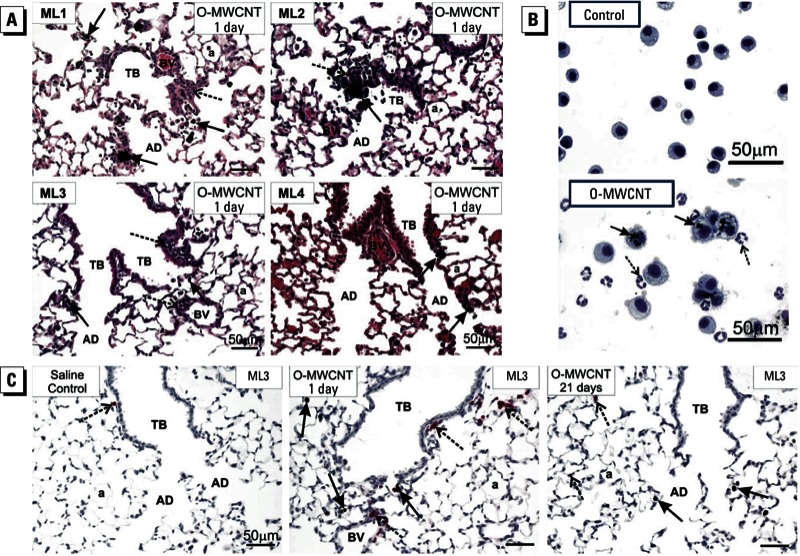
Figure 4.

Results of lung histopathology (A), BAL cytospins (B), and immunohistochemistry (C) for mice exposed to DM only (control) or O-MWCNTs (40 μg/50 μL). Abbreviations: a, alveolus; AD, alveolar ducts; BV, blood vessel; TB, terminal bronchioles. (A) Histopathology showing lung inflammatory response to O-MWCNTs. Centriacinar bronchiolitis/alveolitis (dashed arrows) was induced by O-MWCNTs in three of four labs (ML1, ML2, ML3); one lab (ML4) found some deposition of O-MWCNTs in alveolar ducts with marginal inflammation. Solid arrows indicate macrophages containing O-MWCNT. (B) BAL cytospin images of cells from the lungs of mice exposed to DM (control) or O-MWCNTs; > 95% of macrophages from O-MWCNT–exposed animals were enlarged, activated alveolar macrophages with numerous MWCNT inclusions (solid arrows) and neutrophils that do not contain MWCNTs (dashed arrows). The images are from a single lab (ML3) but are typical of responses from ML1 and ML2. (C) Immunohistochemistry using a monoclonal rat anti-mouse neutrophil (allotypic marker clone 7/4) antibody showing the location of neutrophils (dashed arrows) near terminal bronchioles and in relation to macrophages containing O-MWCNT (solid arrows). Representative images are from ML3.
