


Figure 3.
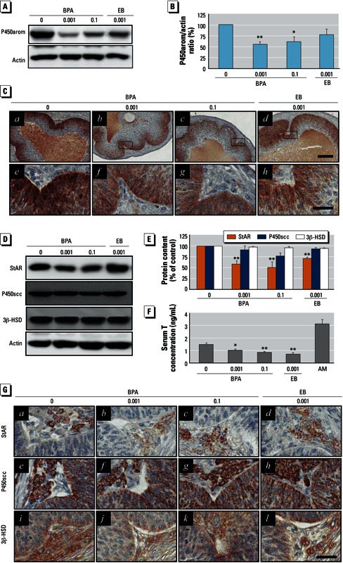
Effect of BPA exposure on P450arom in granulosa cells and StAR, P450scc, and 3β-HSD in the T-I cells of ovarian follicles. Adult female rats were administered BPA (0, 0.001, or 0.1 mg/kg BWU/day) or EB (0.001 mg/kg BW/day) for 90 days by gavage. (A) Western blot analysis for P450arom. (B) Densitometric quantification of the P450arom protein level in isolated granulosa cell protein extracts. (C) Immunohistochemical localization of P450arom in the granulosa cell layers of the large antral follicles; e–h are enlargements of the regions marked in a–d (original magnification: a–d, 100×; e–h, 400×; bars = 80 μm in a–d and 30 μm in e–h). (D) Western blot analysis for StAR, P450scc, and 3β-HSD proteins. (E) Densitometric quantification of StAR, P450scc, and 3β-HSD protein levels in residual ovaries. (F) Serum T levels (mean ± SD) measured by ELISA (n = 12); AM represents adult male serum (positive control). (G) Immunohistochemical localization of StAR, P450scc, and 3β-HSD in T-I layers (original magnification: 400×; bar = 30 μm). For B and E, data represent the mean ± SD of at least three independent experiments. *p < 0.05, and **p < 0.01 compared with control (0 mg/kg BW).