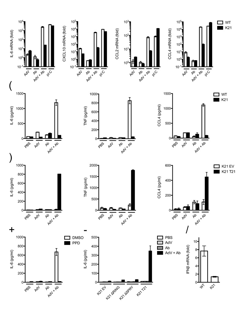
Figure 4. TRIM21 signaling initiates production of proinflammatory cytokines.
(a) Quantitative RT-PCR showing induction of cytokine transcripts after challenge with human serum IgG (Ab), AdV, AdV + Ab or poly I:C in wild-type (WT) or Trim21-deficient (K21) MEFs. Data are represented as fold change above PBS-treated control at 7 h post-infection. (b) ELISAs showing concentration of cytokine protein in wild-type and Trim21-deficient MEF supernatant 72 h post-challenge with indicated treatment. (c) ELISAs showing concentration of cytokines produced by Trim21-deficient expressing empty vector (K21 EV) or human TRIM21 (K21 T21) 72 h post-challenge. (d) ELISA showing concentration of IL-6 of wild-type MEF cell supernatant incubated with DMSO or panepoxydone (PPD) 72 h after challenge with indicated treatment. (e) ELISA showing concentration of IL-6 in supernatant of Trim21-deficient MEF expressing the indicated constructs or transduced with empty vector 72 h post-challenge. (f) Quantitative RT-PCR showing induction of IFN-β transcripts in wild-type and Trim21-deficient MEFs by AdV + Ab over AdV only. For all panels error bars represent SEM from three replicates.

