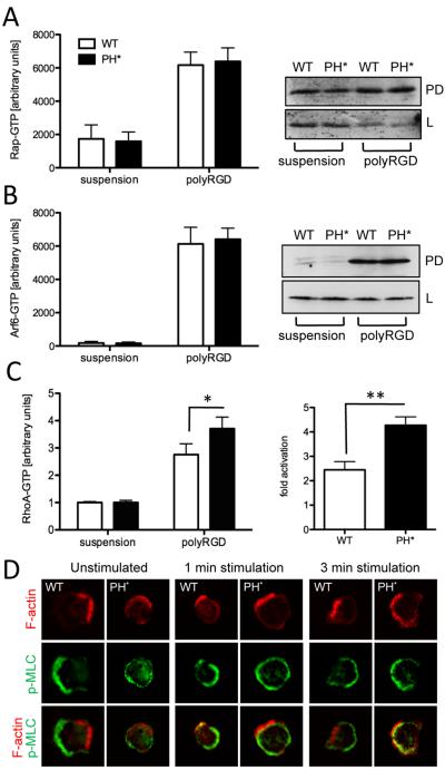
Figure 6. RhoA activation is affected in Arap3PH*/PH* neutrophils.
(A-C) Wild-type control (WT) and Arap3PH*/PH* (PH*) neutrophils were kept in suspension or plated onto polyRGD-coated tissue culture plastic. Cells were lysed with ice-cold lysis buffer. (A, B) Clarified lysates were subjected to ‘pull-down’ assays using GST-Ral GDS as bait to determine GTP-loaded fractions of Rap (A) and using GST-MT2 bait to determine GTP-loaded Arf6 (B). Results obtained from a minimum of five independent experiments were pooled and plotted (mean ± SEM, left); representative examples are shown (right). L, lysates, PD, pull-downs. Blots were probed with an antibody specific for Rap1 (A) and Arf6 (B). (C) Clarified lysates were employed in RhoA G-LISA assays to determine GTP-loaded RhoA. Results from five pooled, independent experiments are presented (mean ± SEM). (A-C) Raw data were analysed by paired T-tests. * p<0.05; ** p<0.001. (D) Indirect assessment of localisation of RhoA activation. Neutrophils were or were not stimulated for the indicated time with 1μM fMLF in solution, allowed to settle on a glass coverslip for 3 minutes, fixed and stained using phalloidin, to visualise filamentous actin and anti phospho myosin light chain. Representative cells from three independent experiments are shown.

