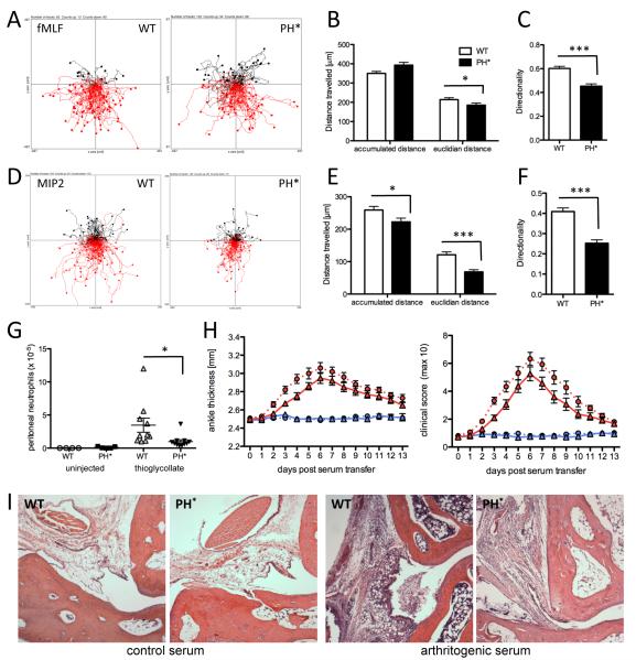
Figure 7. Arap3PH*/PH* neutrophils have a chemotaxis defect.
(A-F) In vitro chemotaxis assays. Bone marrow derived Arap3PH*/PH* and control neutrophils were allowed to chemotax towards 300nM fMLF (A-C) or towards 10nM MIP2 (D-F) in Dunn chambers. Movements were recorded by time-lapse imaging. (A,D) Pooled tracks of individual cells from experiments carried out with three separate cell preparations were plotted using the Ibidi chemotaxis tool plug-in into ImageJ. The source of chemoattractant is at the below. The tracks were analysed using the Ibidi chemotaxis tool’s statistics features. Accumulated and Euclidian distances and directionality are plotted (mean ± SEM; B,C; E,F). (G) Neutrophil recruitment to the peritoneum in a model for sterile peritonitis. Bone marrow chimeras (generated with four bone marrow donors per genotype) were intraperitoneally injected with 0.25ml thioglycollate containing broth. Mice were sacrificed 4.5 hours after injection, their peritonea were flushed and Mac1high GR1-positive neutrophils were counted. Pooled results obtained from two separate experiments are plotted. (B-G) Data were analysed by T-tests (Mann Whitney). * p<0.05; *** p<0.001. (H-I) Serum transfer arthritis. Twelve wild-type and 13 Arap3PH*/PH* bone marrow chimeras were injected with 150μl arthritic serum and six wild-type and six Arap3PH*/PH* bone marrow chimeras were injected with 150μl control serum in two separate experiments. Joints were scored daily for two weeks. Ankle thickness and clinical score are plotted. Circles, and dotted lines, wild-type bone marrow chimeras; triangles and full lines, Arap3PH*/PH* bone marrow chimeras. Blue symbols, control serum; red symbols, arthritogenic serum. The area under the graph was compared by T-test (Mann Whitney); ankle thickness, p=0.053; clinical score, p=0.023. (I) Wax sections of decalcified joints from chimeras reconstituted with control (WT) or Arap3PH*/PH* (PH*) bone marrows induced as indicated were H&E stained to visualise leukocyte infiltration on day 4 after serum injection. Representative examples from sections obtained with 6 arthritogenic and 2 control serum-injected mice mice in two independent experiments are shown.

