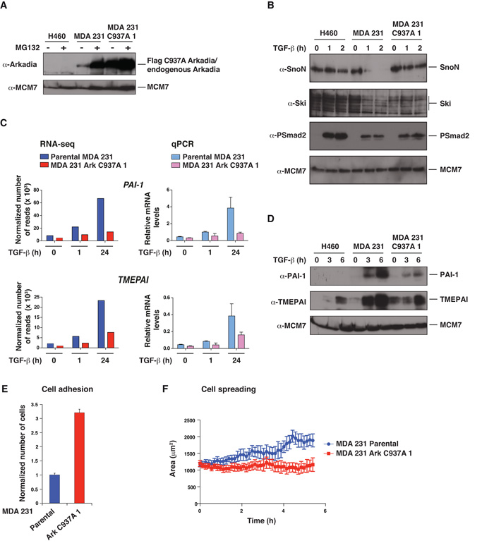
Figure 3. Expression of Arkadia C937A abolishes Arkadia function in MDA-MB-231 cells and alters their adhesion and spreading properties.
A–D. NCI-H460 cells, parental MDA-MB-231 cells, and a stable MDA-MB-231 cell line expressing FLAG-tagged Arkadia C937A (clone 1) were treated ± MG132 for 4 h (A) or with TGF-β for the times indicated (B–D). Whole cell extracts were Western blotted using the antibodies indicated (A, B and D). In C, levels of mRNA for the genes shown were analyzed either by RNA-seq or by qPCR which was normalized to GAPDH. E. GFP-labeled parental MDA-MB-231 cells and Arkadia C937A cells (clone 1) were mixed with an equal number of parental cells expressing mCherry, seeded on a monolayer of HUVEC cells and incubated for 1 h, after which they were washed, fixed and counted. The mean ratio of GFP:mCherry cells and standard deviations from 6 wells is plotted. F. GFP-labeled parental MDA-MB-231 cells or Arkadia C937A cells (clone 1) were seeded on a layer of HUVEC cells, and movies were recorded. Cell spreading was measured by quantification of changes in the area of the cells over time using the Imaris software. The graphs show the means of the average cell area for 4 independent wells for each cell type with standard errors. Similar results were obtained in three independent experiments.

