
Figure 6. Low-level antigen persistence in Ad-LacZ immunized B6 mice.
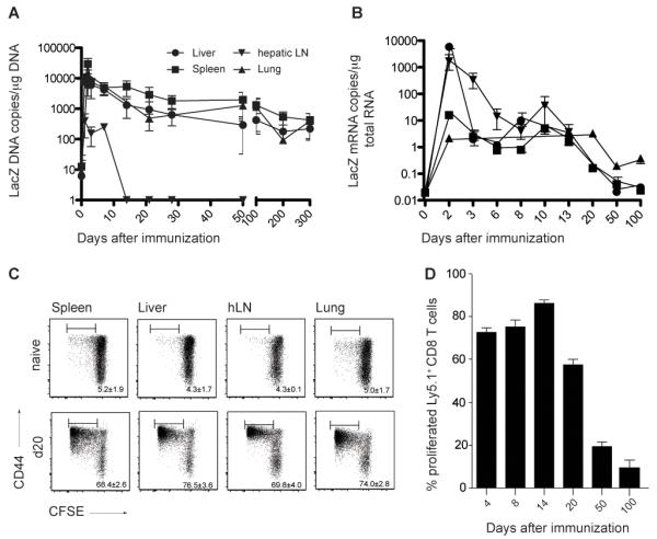
(A) Viral genome distribution after immunization with Ad-LacZ. LacZ DNA copy numbers per μg total DNA were determined with quantitative realtime PCR on different time points after immunization in liver, lung, spleen and hepatic lymph nodes. Values < 10 copies are detectable but not quantifiable. Pooled data from two independent experiments for each time point are shown (Mean±SEM, liver n=3-12, spleen n=3-12, lung n=3-8, hepatic LN n=3-6). (B) βgal expression after immunization with Ad-LacZ. LacZ mRNA copy numbers per μg total mRNA were determined with quantitative realtime PCR at different timepoints after immunization in liver, lung, spleen and hepatic lymph nodes. mRNA copy numbers < 10 are detectable but not quantifiable. Pooled data from two independent experiments for each time point are shown (Mean±SEM, liver n=3-7, spleen n=3-7, lung n=3-6, hepatic LN n=3-4). (C) Low level antigen persistence after i.v. immunization of B6 mice with Ad-LacZ. CFSE-labeled, βgal96-specific, Ly5.1+ TCR-transgenic CD8+ T cells from Bg1 mice transferred on day 20 after Ad-LacZ immunization proliferated in spleen liver, hepatic LNs and lung 3 days after transfer. The numbers indicate the percentage of proliferated Ly5.1+ CD8+ T cells that are donor derived. Pooled data from two independent experiments for each time point are shown (±SEM, spleen n=4, liver n=4, hLN n=4 lung n=4). (D) Percentage of proliferated Ly5.1+ TCR-transgenic CD8+ T cells in spleen analyzed 3 days after adoptive transfer in mice previously immunized with Ad-LacZ on different time points (d4, d8, d14, d20, d50 and d100). Mean percentage (±SEM n=3-5) is indicated.